
Bilateral Tibial Fracture Repair with External Skeletal Fixation in a Dog
A detailed look into a successful treatment at MaxPetZ.
Background
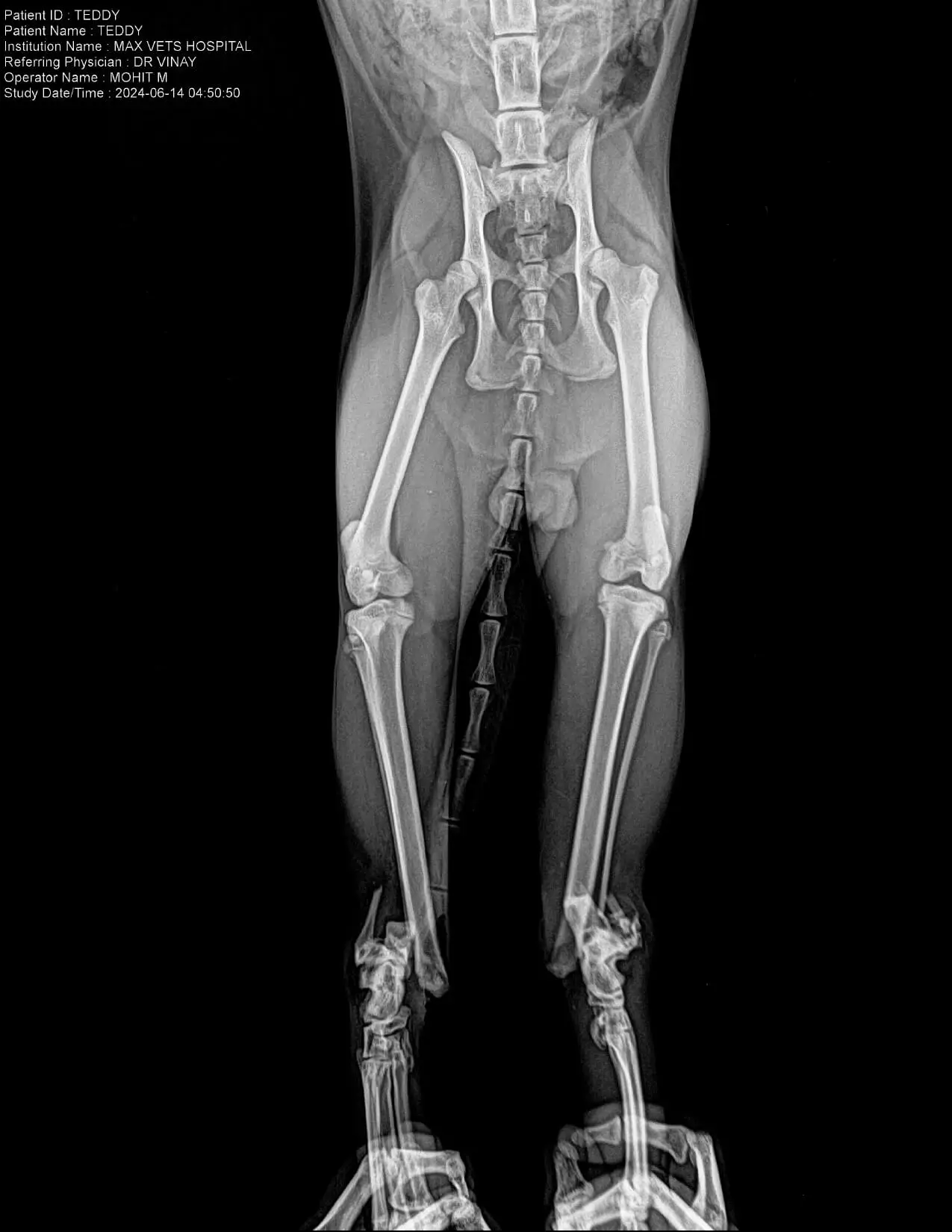
Teddy, a canine patient, with a history of falling from a height the previous day. Initial assessment revealed bilateral tibial fractures—an open fracture in the right hind limb and a closed fracture in the left hind limb. The pet had been given incomplete bandaging at an outside clinic, which Teddy subsequently removed, resulting in exposure of the injured areas.
Initial Presentation & Stabilization
On arrival, Teddy was inappetent, had not taken any fluids since the morning, and showed discomfort in both hind limbs. Physical examination confirmed bilateral distal tibial fractures, with intact deep pain sensation and withdrawal reflexes.
"MRI + CT + 3D planning for safer surgery."
Immediate management included:
- Intravenous fluid therapy for stabilization
- Pain management and anti-inflammatory medications.
- Robert Jones bandaging to immobilize the limbs.
- Owner counseling regarding prognosis, treatment options, and surgical requirements.
Diagnostic Workup
Over the next few days, Teddy underwent:
- CBC, LFT, and KFT blood panels to assess systemic health and anesthetic risk.
- X-rays confirming fracture configuration.
- AFAST and TFAST ultrasonography to evaluate internal organ health and rule out thoracic/abdominal trauma.
Findings revealed elevated liver enzymes and bilirubin, necessitating supportive hepatic therapy before proceeding with surgery.
Surgical Intervention
On once Teddy’s systemic parameters improved, external skeletal fixation (ex-fixation) was performed under general anesthesia.
- Two cross pins were placed from the tibia to the tarsals in each limb for stabilization.
- Muscles and subcutaneous tissues were closed with absorbable sutures, and skin with non-absorbable sutures.
- Postoperative recovery from anesthesia was smooth.
Postoperative Care & Rehabilitation
Teddy was managed with:
- Injectable antibiotics, analgesics, and anti-inflammatories for the first three days post-op.
- Oral medications for pain, joint support, and bone healing over the following weeks.
- Regular bandage changes every three days.
- E-collar application to prevent interference with surgical sites.
- Scheduled follow-ups for suture removal, pin site inspection, and physiotherapy guidance.
Follow-Up & Implant Removal
Over the next two months, Teddy attended regular reviews. Radiographic assessment confirmed satisfactory healing. On August 17 and 26, 2024, staged implant removal procedures were carried out successfully, with no complications noted.
Outcome
By the end of the treatment period, Teddy was active, bearing weight on all limbs, and showing a good range of motion. The fractures had healed satisfactorily, and the patient’s quality of life had returned to normal.
Discussion
This case highlights:
- The importance of timely stabilization in open fractures to prevent infection and further trauma.
- Preoperative optimization, especially in patients with concurrent systemic issues such as elevated liver enzymes.
- External skeletal fixation as an effective technique for stabilizing complex, bilateral hind limb fractures in small breed dogs.
The need for owner compliance in post-operative care for successful outcomes.
Treatment Timeline
Day 1
Initial Trauma
Teddy fell from a height, sustaining bilateral tibial fractures (open fracture in right hind limb, closed fracture in left hind limb).
Day 1
Initial Stabilization
IV fluids, pain management, anti-inflammatories, and Robert Jones bandaging applied. Owner counseled on prognosis and surgical options.
Days 2–3
Diagnostic Workup
CBC, LFT, KFT, X-rays, and ultrasonography performed. Elevated liver enzymes and bilirubin detected; hepatic therapy initiated before surgery.
After stabilization
Surgical Intervention
External skeletal fixation performed under anesthesia with two cross pins placed in each tibia-to-tarsal segment. Muscles and skin sutured; smooth anesthesia recovery.
First 3 days
Postoperative Care
Injectable antibiotics, analgesics, and anti-inflammatories administered; oral medications continued for bone healing and joint support.
Weeks 1–8
Rehabilitation
Regular bandage changes, E-collar to protect surgical sites, physiotherapy guidance, and suture removal visits.
August 17 & 26, 2024
Implant Removal
Staged removal of external fixation implants performed successfully; radiographs confirmed good fracture healing.
Post-treatment
Recovery Outcome
Teddy active, weight-bearing on all limbs, with full range of motion and return to normal quality of life.
Your Pet Deserves Specialist Care
If you're concerned about your pet's health, our team is ready to provide the advanced care and compassion they need.