
Successful Brain Tumor Surgery in a Canine Patient
A detailed look into a successful treatment at MaxPetZ.
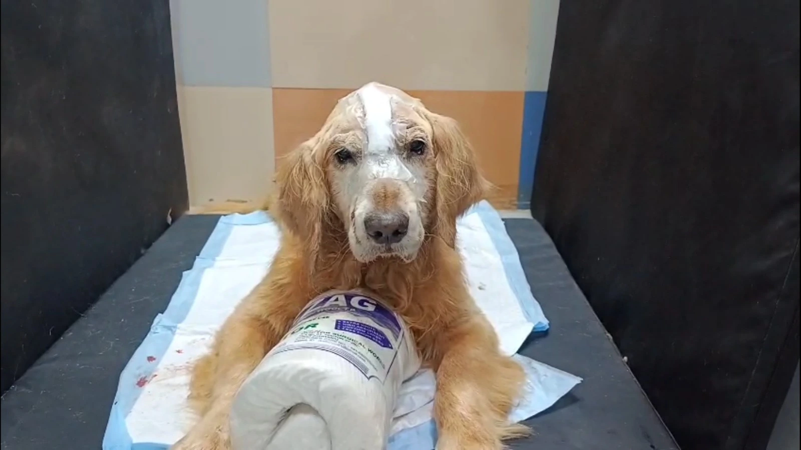
Background
Juno, a canine patient, was referred to MaxPetZ with a history of seizures and noticeable behavioral changes. The episodes had increased in frequency, prompting advanced neurological investigation. A CT scan revealed a solitary mass in the frontal lobe of the brain, measuring approximately 24 x 28.6 mm.
Pre-Surgical Assessment and Planning
Before surgery, Juno underwent a thorough evaluation, including:
Comprehensive blood work to assess overall systemic health.
Liver and kidney function tests to evaluate anesthetic risk.
Cardiac evaluation to ensure cardiovascular stability under anesthesia.
Supportive management to control intracranial pressure and stabilize neurological function prior to intervention.
To ensure maximal surgical precision, the MaxPetZ team employed 3D printing technology. Using Juno’s CT data, a custom 3D model of her skull was created, enabling the surgeons to identify anatomical landmarks in advance. This model allowed the team to plan the craniotomy approach in detail, minimize surgical time, and reduce intraoperative risks.
Surgical Procedure
On January 5, 2025, the surgical team performed a transfrontal craniotomy under general anesthesia.
Key steps included:
Aseptic preparation and a midline incision over the skull from the medial canthi to the bregma.
Careful separation of subcutaneous tissues and musculature to expose the frontal bone.
Removal of a triangular bone section (prude diamond craniotomy) to access the frontal sinus and underlying dura.
Visualization of the dura and meticulous tumor excision using a scooping technique until only healthy brain tissue was visible.
Replacement and secure fixation of the bone plate using a four-hole technique.
Layered closure of muscles and skin in a routine fashion.
The excised tissue was immediately preserved and sent for histopathological analysis to confirm the tumor type and ensure complete removal.
Postoperative Care
Following surgery, Juno was admitted to the Intensive Care Unit for continuous monitoring and specialist nursing care. Postoperative management included:
Pain control via fentanyl transdermal patch for sustained analgesia.
Anti-inflammatory therapy (including corticosteroids) to minimize cerebral edema.
Anticonvulsant medication to reduce seizure risk during recovery.
Gradual transition to oral medications for long-term neurological support.
Calm, low-stimulation environment to support neurological healing.
Recovery and Follow-Up
In the days following surgery, Juno demonstrated significant improvement:
She became progressively more alert, responsive, and active.
Appetite returned, and her seizure frequency reduced markedly.
Motor coordination improved, and she was able to interact normally with her owners.
Sutures were removed on January 16, 2025, by which time Juno was in good condition, had gained weight, and showed sustained neurological stability.
Outcome
The surgery successfully achieved complete tumor removal, as confirmed by histopathology. Juno’s quality of life improved significantly, with seizure control, restored activity levels, and an excellent recovery profile. Follow-up medications were prescribed to manage inflammation, maintain neurological stability, and prevent recurrence of symptoms.
Discussion
This case highlights several key aspects of modern veterinary neurosurgery:
Advanced Imaging: High-resolution CT scanning allowed precise localization of the tumor, guiding surgical planning.
3D Printed Surgical Models: The use of a patient-specific skull model enabled accurate identification of surgical landmarks, minimized operative risks, and optimized surgical efficiency.
Specialist Surgical Expertise: The skilled performance of a transfrontal craniotomy ensured complete tumor removal while preserving healthy brain tissue.
Postoperative Management: Intensive care, pain control, anti-inflammatory therapy, and anticonvulsant use played crucial roles in Juno’s recovery.
Owner Compliance: Adherence to follow-up schedules and home care instructions contributed greatly to the long-term success of the procedure.
Treatment Timeline
Day 1
Initial Presentation
Dog presented with seizures and behavioral changes; referred to MaxPetZ Neurology.
Day 1
Diagnostic Imaging (CT)
CT scan identified a solitary frontal lobe mass (~24 x 28.6 mm).
Days 2-3
Pre-Surgical Assessment
Comprehensive blood work; liver/kidney function; cardiac evaluation for anesthesia risk.
Days 2-4
Stabilization & ICP Management
Supportive therapy to control intracranial pressure and optimize neurological status.
Days 3-5
3D Printing & Surgical Planning
Patient-specific skull model printed from CT data; craniotomy approach rehearsed to reduce intraoperative risk.
Surgery Day
Anesthesia & Aseptic Prep
General anesthesia induced; midline scalp incision prepared under sterile conditions.
Surgery Day
Transfrontal Craniotomy
Triangular bone flap (prude diamond) created; dura exposed and tumor excised until healthy tissue visualized; bone plate fixed (four-hole technique); layered closure.
Surgery Day
Specimen Handling
Excised tissue preserved and dispatched for histopathology.
Days 1-3 Post-op
ICU Postoperative Care
Continuous monitoring; fentanyl patch for analgesia; corticosteroids for edema; anticonvulsants for seizure control.
Days 4-6 Post-op
Transition & Discharge
Gradual switch to oral medications; low-stimulation environment; discharge with home-care instructions.
Days 7-10 Post-op
Early Recovery
Improved alertness, appetite, motor coordination; marked reduction in seizure frequency.
Around Day 11 Post-op
Suture Removal & Review
Sutures removed; patient in good condition with weight gain and stable neurology; histopathology confirms complete tumor removal.
Ongoing
Long-term Management
Continue prescribed anti-inflammatory and anticonvulsant regimen; periodic neurological follow-ups to monitor for recurrence.
Your Pet Deserves Specialist Care
If you're concerned about your pet's health, our team is ready to provide the advanced care and compassion they need.