
Total Hip Replacement for Severe Hip Dysplasia in a Golden Retriever
A detailed look into a successful treatment at MaxPetZ.
Background / Introduction / Case Presentation
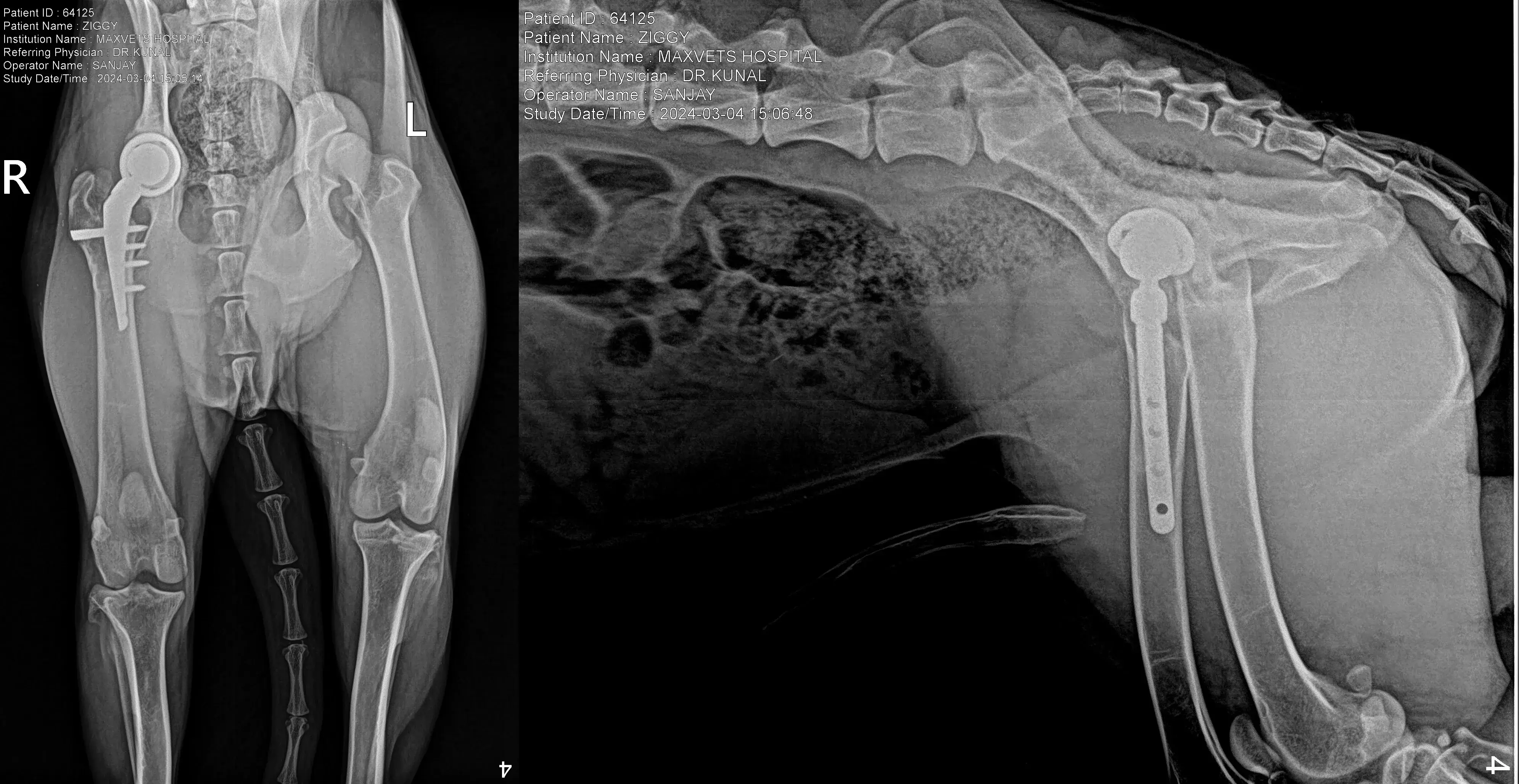
Simba, a 2-year-old male Golden Retriever weighing 34 kg, was admitted to MaxPetZ on July 19th, 2025, with a primary diagnosis of severe hip dysplasia in the left hip joint. He presented with a six-month history of hind limb lameness, stiffness upon rising, reluctance to jump or climb stairs, and reduced activity levels. Previous medical management, including NSAIDs, joint supplements, and controlled exercise, had failed to provide sustained relief. Total Hip Replacement (THR) is a procedure designed to relieve pain and restore function for dogs with severe hip conditions, often indicated for hip dysplasia in large breeds and end-stage osteoarthritis. At MaxPetZ, THR is generally preferred over Femoral Head Osteotomy (FHO) due to superior outcomes and improved quality of life, with over 95% of dogs expected to enjoy an active, pain-free lifestyle.
Clinical Findings / Orthopedic Examination
Upon orthopedic examination, Simba exhibited pain and crepitus on manipulation of the coxo-femoral joint, decreased range of motion, and muscle atrophy of the gluteal and thigh muscles. (Note: The source specifically mentions the right coxo-femoral joint in the examination findings, while the diagnosis and procedure were for the left hip). Dogs undergoing evaluation for THR are typically assessed for gait, posture, pain, and joint stability using tests like Ortolani, Barlow’s, and Barden’s tests. A positive Ortolani sign or a reducible hip is part of the surgical candidacy checklist.
Diagnostics / Diagnostic Work-Up / Imaging
- Diagnostic Imaging: Pelvic radiographs (ventrodorsal extended hip view) confirmed severe degenerative joint disease (DJD) of the left hip joint, characterized by loss of joint space, osteophyte formation, femoral head remodeling, and acetabular sclerosis. The contralateral hip showed mild to moderate DJD but remained functional. Radiographic views, including VD pelvic, frog-leg, yoga, medio-lateral femur, and lateral ileum, are crucial for evaluating hip structure and laxity, often involving measurements like the Distraction Index (DI). A DI greater than 0.7 is a key factor in surgical candidacy.
- Pre-anesthetic Assessment: Simba underwent comprehensive pre-anesthetic testing, with all results within normal limits:
- Complete Blood Count (CBC)
- Liver Function Tests (LFT)
- Kidney Function Tests (KFT)
- Electrolytes
- Coagulation Profile (Normal PT and aPTT)
- Thoracic Radiographs (No evidence of cardiopulmonary disease)
- Electrocardiogram (ECG) (Normal sinus rhythm, no arrhythmias)
- Body Condition Score (BCS): Simba had a BCS of 6/9, indicating a healthy weight, which is favorable as high BMI increases complication risk.
Based on these results, Simba was cleared for general anesthesia and surgery.
Pre-Surgical Preparation / Stabilization
Simba received pre-medication intramuscularly (IM) with Acepromazine (0.02 mg/kg), Morphine (0.3 mg/kg), and Glycopyrrolate (0.01 mg/kg). An intravenous (IV) catheter (20G) was placed in the cephalic vein, and Ringer’s Lactate solution was administered at 5 ml/kg/hr throughout the surgery. Anesthesia was induced intravenously (IV) with Propofol (4 mg/kg to effect). Antibiotic prophylaxis was initiated with Cefazolin (22 mg/kg IV) at induction, repeated every 90 minutes intraoperatively.
Surgical Procedure / Surgical Management
The Total Hip Replacement for Simba was performed on July 19th, 2025, using the Prime Hip Implant System for the left hip.
- Maintenance: Anesthesia was maintained with Isoflurane in 100% oxygen via a rebreathing circuit, with Intermittent Positive Pressure Ventilation (IPPV). Continuous monitoring included ECG, SpO₂, EtCO₂, temperature, and Blood Pressure (BP).
- Intraoperative Analgesia: Fentanyl Constant Rate Infusion (CRI) (5 µg/kg/hr) was administered, along with an epidural of morphine (0.1 mg/kg) and bupivacaine (0.5 mg/kg) for perioperative analgesia.
- Temperature Management: Intraoperative temperature was maintained using a circulating warm water blanket and warm IV fluids.
- Approach: A standard craniolateral approach to the hip joint was utilized.
- Femoral Head Resection: The femoral head was removed using an oscillating saw at the anatomical neck.
- Acetabular Preparation: Sequential reaming was performed to remove diseased cartilage and create a congruent hemispherical bed. A cementless press-fit acetabular cup was then inserted, achieving excellent primary stability.
- Femur Preparation: Sequential broaching was done to fit the Prime Hip femoral barrel plate (3.5mm). Titanium 3.5mm screws were inserted in a monocortical manner proximally (bilaterally) and distally. The Prime Hip system is designed with a lateral locking plate and double-locking bolt, which avoids intramedullary canal preparation, preserving endosteal blood supply and reducing fracture risk. This system also features a pre-angled barrel at 135 degrees and a hydroxyapatite-coated barrel to support osteointegration.
- Implant Insertion: The barrel plate (cementless, titanium alloy) was fixed for the lateral locking bolt, and the femoral head was attached to the bolt.
- Final Reduction: Final reduction was achieved, demonstrating full stability in extension, flexion, and rotation without any luxation tendency.
- Closure: Routine layered closure was performed using absorbable sutures for fascia, subcutaneous tissue, and intradermal skin layers. A sterile dressing was applied.
- Intraoperative Details: The procedure lasted 95 minutes, with approximately 80 ml of blood loss, and no transfusion was required. Crucially, no intraoperative complications were noted.
Postoperative Care / Postoperative Management
Hospitalization (July 19th – July 23rd, 2025):
- Day 0 (Surgery Day): Simba had a smooth recovery and was extubated uneventfully. He was monitored for hypothermia, hypoventilation, and pain. IV fluids were continued for 12 hours post-op, and Fentanyl CRI was maintained for 12 hours before transitioning to multimodal oral analgesia.
- Day 1 Post-op: Simba was bright, alert, and eating a soft diet. He exhibited minimal but present weight-bearing on the operated limb with sling support. The wound was clean and dry with no swelling. Cryotherapy was applied 3–4 times/day to the surgical site, and passive range-of-motion exercises were initiated.
- Day 2 Post-op: Gradual improvement in comfort and limb use was observed. Sling-assisted short walks (2–3 minutes, 4 times/day) were introduced. The surgical site was checked, showing no discharge and only mild expected bruising. Antibiotics and NSAIDs were continued.
Medications at Discharge:
- Carprofen: 4 mg/kg orally once daily for 10 days
- Tramadol: 3 mg/kg orally every 8–12 hours for 7 days
- Cefpodoxime: 10 mg/kg orally once daily for 7 days
- Omeprazole (Gastroprotectant): 1 mg/kg orally once daily for 10 days
Post-operative Home Care Instructions:
- Activity Restriction: Strict rest for the first 6 weeks. Walks only with sling support for toileting, 3–4 times/day. No running, jumping, or stairs.
- Physiotherapy: Passive range-of-motion exercises 3–4 times/day from day 3 onward. Hydrotherapy can be considered after suture removal if healing is optimal. This aligns with rehabilitation goals of promoting bone ingrowth, encouraging normal limb use, and minimizing muscle atrophy.
- Incision Care: Keep clean and dry. Prevent licking/chewing using an Elizabethan collar. Monitor for swelling, redness, discharge, or foul odor.
- Bandage/Dressing: Remove in 48 hours if no discharge.
- Ice Packs: 10–15 minute sessions, 3–4 times/day for the first 3 days, then switch to warm compresses.
Recovery & Follow-Up / Recovery Timeline
The rehabilitation protocol is structured in phases to optimize long-term outcomes, focusing on soft tissue healing, progressive strengthening, and gradual activity resumption.
- Immediate Postoperative Phase (0–14 Days): Simba was confined to a crate or small room with non-slip flooring and had brief lead walks for basic needs. Pain management was provided, and passive exercises initiated.
- Day 10–12: Follow-up for suture removal and wound assessment.
- Controlled Recovery Phase (2–8 Weeks): Supervised lead walks gradually increase in duration. Hydrotherapy may be introduced after incision healing, and carpeted stairs are permitted at six weeks. High-impact activity is restricted.
- Week 6: Recheck radiographs to assess implant position and bone healing. Annual monitoring with regular imaging, gait videos, blood work, and owner surveys is recommended to detect issues early.
- Advanced Functional Recovery (8–12 Weeks): Gradual introduction of controlled off-leash activity, varied surfaces, simple agility tasks, and strengthening exercises. Radiographic follow-up at ten weeks.
- Month 3: Final recheck, with gradual return to normal activity if healing is complete.
- Long-Term Return to Normal Function (>12 Weeks): Most dogs are allowed unrestricted activity by 12–14 weeks, with an extended ramp-up for working/sporting animals. Repetitive high-impact actions should be avoided.
Outcome / Prognosis
Simba's prognosis for significant improvement in mobility and pain reduction is excellent, given the advanced osteoarthritis and the stability achieved intraoperatively, provided there is proper post-operative rehabilitation and lifelong joint care. Factors like early gait restoration, compliance with rehabilitation, and absence of complications are favorable indicators for implant longevity. MaxPetZ clinical data for Prime Hip systems shows a 96.1% survival rate, with low rates of dislocation (2.8%), infection (1.3%), and loosening (1.3%).
Discussion
This case highlights the successful application of the Prime Hip system for canine THR in a young Golden Retriever with severe hip dysplasia. The Prime Hip system, which utilizes a lateral locking plate and double-locking bolt mechanism, offers several advantages over traditional intramedullary fixation systems. It eliminates the need for intramedullary canal preparation, thereby preserving endosteal blood supply and reducing the risk of femoral fractures. Its adaptability to femoral variations like coxa vara/valga and genu valgum, along with modular bolt lengths, ensures greater stability and functional outcomes.
The detailed pre-operative assessment and adherence to a strict anesthetic and surgical protocol minimized intraoperative risks, as evidenced by Simba having no intraoperative complications. Post-operative rehabilitation, including strict activity restriction, passive range-of-motion exercises, and gradual activity progression, is crucial for promoting bone ingrowth and preventing complications like dislocation or muscle atrophy. The success rate of THR, particularly with modern systems like Prime Hip, far surpasses that of Femoral Head Ostectomy (FHO). FHO is considered a "rescue" procedure by MaxPetZ and is associated with unpredictable pain relief, limb shortening, restricted hip movement, and muscle atrophy, offering less predictable outcomes, especially in larger dogs. The superior biomechanical function and pain relief offered by THR make it the preferred first-line surgical treatment. Long-term monitoring, including regular radiographs and clinical evaluations, is essential to assess implant stability and function, ensuring continued positive outcomes.
Conclusion
Simba's case demonstrates an excellent prognosis for restored mobility and a pain-free life following a successful Total Hip Replacement with the Prime Hip system, emphasizing the importance of advanced surgical techniques, meticulous perioperative care, and diligent post-operative rehabilitation.
Treatment Timeline
Pre-surgery
Initial Presentation & History
Six-month history of hind limb lameness, stiffness on rising, reluctance to jump; medical management failed.
Pre-surgery
Diagnostics & Candidacy
Pelvic radiographs confirmed severe DJD of the left hip; CBC, LFT, KFT, electrolytes, coagulation profile, thoracic radiographs, and ECG within acceptable limits; BCS 6/9.
Day 0 (July 19, 2025 – morning)
Pre-Anesthetic Preparation
Premed IM (acepromazine, morphine, glycopyrrolate); IV catheter placed; cefazolin started; fluids RL 5 ml/kg/hr.
Day 0
Anesthesia Induction & Maintenance
Induced with propofol; maintained on isoflurane with IPPV; continuous monitoring (ECG, SpO₂, EtCO₂, temperature, BP); fentanyl CRI plus epidural morphine and bupivacaine.
Day 0 (July 19, 2025)
Surgery — Total Hip Replacement (Left)
Prime Hip system used via craniolateral approach; acetabular reaming and cementless cup placement; femoral barrel plate (3.5 mm) with locking screws; lateral locking bolt and HA-coated barrel; stable reduction achieved; layered closure.
Day 0
Intra-op Summary
Duration ~95 minutes; ~80 ml blood loss; no intraoperative complications.
Day 0 (evening)
Immediate Post-op Recovery
Smooth extubation; IV fluids continued ~12 h; fentanyl CRI maintained then plan to transition to oral multimodal analgesia.
Post-op Day 1 (July 20, 2025)
Hospital Day 1
Bright, alert, eating soft diet; minimal weight-bearing with sling; incision clean/dry; cryotherapy 3–4×/day; passive ROM initiated.
Post-op Day 2 (July 21, 2025)
Hospital Day 2
Improving comfort and limb use; sling-assisted short walks (2–3 min, 4×/day); mild expected bruising; continue antibiotics/NSAIDs.
Post-op Day 3 (July 22, 2025)
Hospital Day 3
Continued stabilization and nursing care; plan discharge next day with home meds and rehab instructions.
July 23, 2025
Discharge
Sent home on carprofen, tramadol, cefpodoxime, omeprazole; strict rest, E-collar, incision care, ice packs 3 days then warm compresses.
Post-op Days 1–14 (at home)
Early Home Care
Crate/small-room rest; sling walks for toileting; passive ROM 3–4×/day; bandage off at 48 h if dry.
Post-op Day 10–12
Suture Removal
Clinic visit for suture removal and wound assessment.
Weeks 2–8
Controlled Recovery
Gradual increase in supervised leash walks; introduce hydrotherapy after incision healing; stairs permitted around week 6; high-impact activity restricted.
Week 6
Six-Week Recheck
Radiographs to evaluate implant position and bone ingrowth; adjust rehab plan as needed.
Weeks 8–12
Advanced Functional Recovery
Begin controlled off-leash activity, surface variation, simple strengthening/agility; radiographic follow-up around week 10.
Month 3
Three-Month Recheck
Clinical and radiographic evaluation; gradual return to normal activity if healing complete.
≥12–14 weeks and ongoing
Long-Term Return to Function
Most dogs resume unrestricted activity by 12–14 weeks; continue lifelong joint care and periodic monitoring to optimize implant longevity.
Your Pet Deserves Specialist Care
If you're concerned about your pet's health, our team is ready to provide the advanced care and compassion they need.